St Petersburg University scientists propose novel method to assess organ viability during abdominal surgery
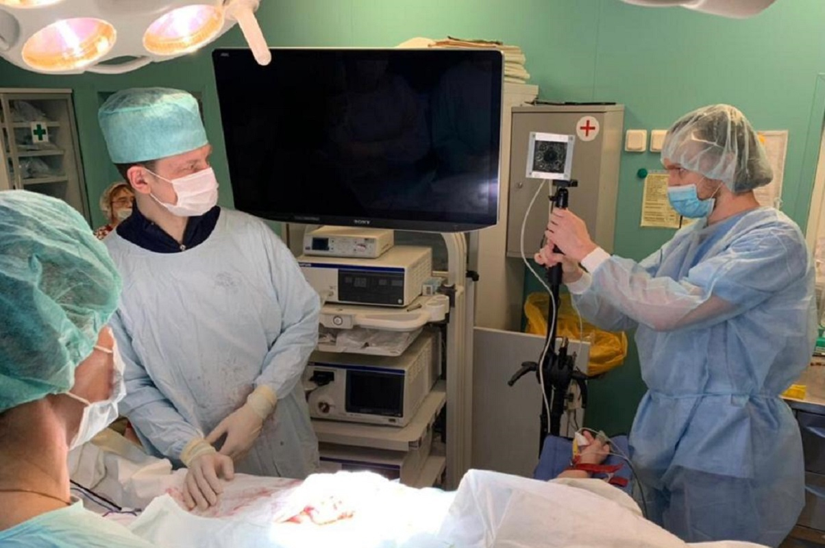
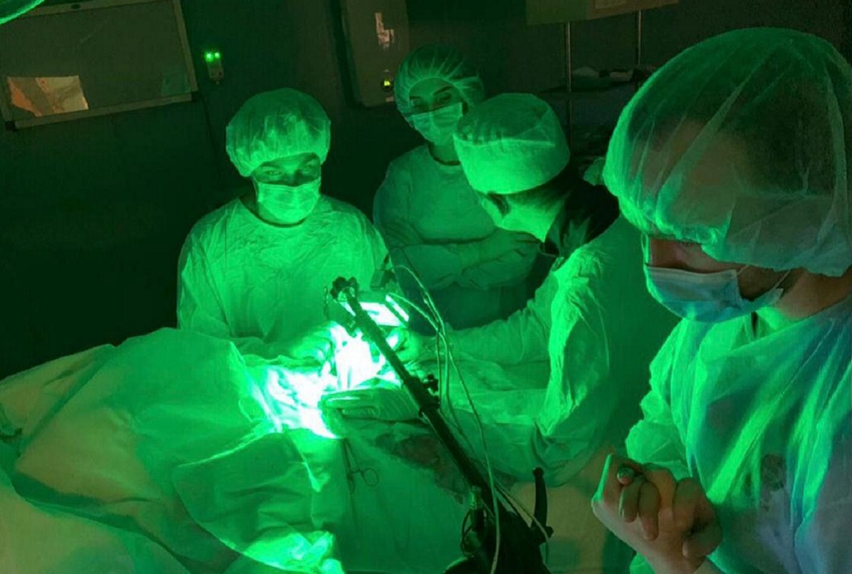
Russian scientists have introduced into clinical practice an alternative method for assessing the blood supply of organs and tissues during abdominal surgery. This method, referred to as imaging photoplethysmography, provides noninvasive evaluation of capillary blood flow, using images in green light from a special camera. It can also help to avoid post-operative complications and reduce mortality.
Their paper has been published in the scientific journal Scientific Reports, which is part of Nature, a group of authoritative general scientific journals.
The team of scientists responsible for this breakthrough includes: Professor Viktor Kashchenko, Head of the Department of Faculty Surgery at St Petersburg University and also Deputy Director General for Scientific and Educational Work and Chief Surgeon at the Sokolov Northwestern District Scientific and Clinical Centre; Aleksandr Lodygin, Associate Professor in the Department of Faculty Surgery at St Petersburg University; and Aleksei Kamshilin and Valerii Zaitsev, researchers at the Institute of Automation and Control Processes, which is part of the Far Eastern Branch of the Russian Academy of Science.
An innovative method, photopletysmography could replace traditional fluorescent imaging (ICG) technology. Today, ICG is one of the critical components in ensuring the safety of surgical interventions. With its help, a surgeon can assess how viable organs are and plan an operation correctly, including the amount of tissue that should be removed. This method, however, has a number of limitations: it is difficult to use in cases of liver insufficiency, as it is necessary to inject the patient with a special dye (ICG-indocyanine green) as an intravenous indicator, and to use expensive imported equipment, which is available in far from all health centres.
In essence, this new technique is somewhat reminiscent of assessing saturation with a pulse oximetre, except that photopletysmography is a more complex technology, and capillary blood flow is assessed remotely, using a green-light camera.
Viktor Kashchenko, Chief Surgeon at the Sokolov Northwestern District Scientific and Clinical Centre
It is a specially designed measuring system with a digital electrocardiograph. The images of tissues recorded on the camera are processed using software that makes it possible to assess their blood supply. Unlike other methods, measurements can be carried out any number of times, and it can also be used for dynamic monitoring during surgery. It was put into practice by surgeons at the Sokolov Northwestern District Scientific and Clinical Centre, and used in a paper analysing the clinical cases of 14 patients.
The scientists are also contemplating the possibility of using artificial intelligence to process the data obtained during photopletysmography. The main result that doctors anticipate is a reduced risk of complications after surgery. Above all, surgeons hope to avoid one of the most severe complications: anastomotic leakage after resection or reconstruction of abdominal organs, including in cases of colon cancer or stomach cancer. Today, this complication is encountered, depending on the type of operation, in up to 10% of patients, and it can also lead to death in the post-operative period.
This research was supported by the Russian Science Foundation (project No 21-15-00265).


