Our immune system is both a boon and a bane

St Petersburg University researchers have been studying the aggressive behaviour of the immune system towards its own organism, as exemplified by sarcoidosis. Their research will shed light on the process of this disease and will also improve its diagnosis.
The immune system protects the body against viruses, bacteria and fungi. In some cases, however, its reaction spirals out of control and damages healthy cells and organs. St Petersburg University researchers investigated autoimmune behaviour under a large grant or a ‘megagrant’ to study the causes and mechanisms of autoimmune diseases and the use of immunological tools for their diagnosis and treatment.
The investigation was conducted under megagrant No 14.W03.31.0009 bestowed by the Government of the Russian Federation on February 13, 2017, which provides government support for research to be carried out under the guidance of leading scientists.
As one of the avenues of research, the scientists chose sarcoidosis. This is a non-communicable disease characterised by the appearance of nodular formations in organs and tissues of the body, most commonly in the lungs. The disease triggers inflammatory responses that take a toll on the entire body.
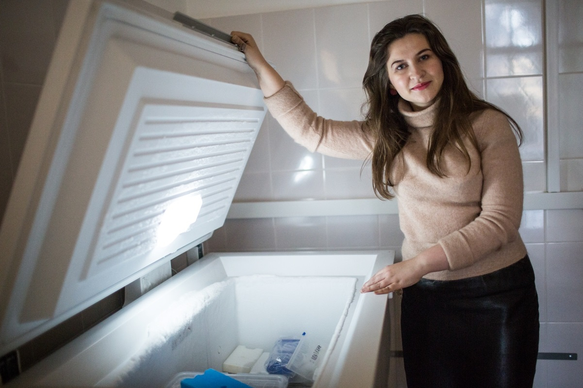
‘There are diseases whose pathogeneses are little-understood,’ relates Nataliia Basantsova, an assistant lecturer in the Department of Faculty Therapy at St Petersburg University and a neurologist at the St Petersburg Research Institute of Phthisiopulmonology. ‘It is obvious that the immune system plays an important role in pulmonary sarcoidosis. Because several of its components are overly active, it damages not only lung tissue but the body as a whole. Among other things, this leads to development of small fibre neuropathy (SFN), and this is what I was invited to carry out research on a year and a half ago.’
The reflective immune system
‘Normally, the immune system works like this: there is a group of cells that very carefully screen each and every foreign body that makes its way into the body. This ensures a prompt response to the intrusion of pathogens,’ the neurologist explains. ‘As long as these cells function properly, the immune system is able to cope with infections. But if they start letting things slip through, tumours and infectious diseases begin to develop, and if, conversely, they become too active, autoimmune diseases evolve.’
Autoimmune responses arise when there is a disbalance in the so-called T-component of the immune system, whose key ‘participants’ are T-helper cells, T-killer cells and T-suppressor cells. T-helpers recognise foreign molecules and send a message to activate other immune cells. T-killers destroy afflicted cells to prevent an infection from spreading further. T-suppressors perform a regulatory function and stifle a superfluous immunological response. ‘In theory, T-cells should protect us, but in the presence of an autoimmune disease their functions go awry: some of them become hyperactive and others, hypoactive,’ Ms Basantsova recounts.
A malfunctioning of T-cells triggers the release of more cytokines than are needed. Cytokines are ‘informational molecules’ that transmit signals between lymphocytes, and they differ according to their characteristics. Some are pro-inflammatory (they cause inflammation), others are anti-inflammatory (they reduce it) and still others are regulatory (they monitor the innate and acquired immune systems). ‘Anybody who has ever come down with the flu has felt the effect of pro-inflammatory cytokines,’ the scientist points out. ‘The virus itself does not cause your temperature to rise and your muscles to ache. It’s the cytokines that are to blame, triggering, as they do, the appropriate response.’
In patients with pulmonary sarcoidosis and other autoimmune diseases, the number of pro-inflammatory cytokines – IL-1β, IL-6, IL-8 and FNO-α – increases. The cytokines damage not only the lung tissue but also the slender nerve fibres, the myelinated neurons of the A-delta type (with a protective sheath of myelin) and the unmyelinated C-type nerve fibres (with no sheath), which all of the body’s internal organs and tissues are infused with. This leads to the appearance of neurological symptoms, including dysrhythmia of the heart and polymyositis (muscle pain) and dermatomyositis (skin rashes), both of which make it hard to sleep at night.
According to Ms Basantsova, nocturnal aggravation of these symptoms is connected with the classical theory of Ronald Melzack and Patrick Wall. It postulates that algesic (pain-causing) and muscular impulses are processed by the same neurons. If, for example, a person cuts their finger, they reflexively start to squeeze the area of the wound. They do this not so much to stop the bleeding as to activate the impulses of the nearby muscles. Since information about the pain and the contractions of the muscle fibres come simultaneously to one and the same centre in the brain, it has to make a choice between them and, for evolutionary reasons, it gives preference to the muscles. So, during the day, when they are on the move or involved in sport, the patient feels fine. But when they go to bed, the muscles relax and the discomfort returns. Hormonal fluctuations also exacerbate the symptoms. ‘It is thought that male patients and women during menopause feel the pain more acutely,’ the researcher notes.
A diagnosis based on a millimetre of skin
Pulmonary sarcoidosis and its neuropathic manifestations are very difficult to diagnose. Detection of autoimmune diseases requires the use of instrumental procedures along with a meticulous physical examination and interrogation by a doctor. In 70 percent of the cases, the disease takes a benign form, and in half of the patients it is discovered during a routine examination. According to rough estimates, there are currently hundreds of millions of people in the world with progressive SFN for various reasons, including sarcoidosis. Perhaps this figure is even higher.
What is more, doctors are often unaware that the complex of autonomic symptoms that go along with sarcoidosis are its neurological manifestation.
Nataliia Basantsova, a neurologist at the St Petersburg Research Institute of Phthisiopulmonology
If, however, a doctor grows suspicious and decides to carry out the standard test, electroneuromyography, it is unlikely that he will be able to confirm a diagnosis. This test discloses irregularities only in large nerves; small fibres are beyond its scope. ‘In order to judge whether or not there is evidence of SFN, you have to go beyond the standard protocol of this procedure,’ says Ms Basantsova, ‘and also possess the required expertise.’
Thus, the ‘gold standard’ for the determination of SFN is a skin biopsy. It makes it possible to assess much more precisely the condition of small nerve fibres in the epidermis. As a rule, this analysis is carried out on the other side of the body, where clinical responses are more apparent. First of all, a specialist anaesthetises the area and treats it with an antiseptic. Next, with a biopsy needle, they take a small slice of skin approximately three or four millimetres in size and fix it in a special solution. After that, they dye it and put it under a microscope.
According to Ms Basantsova, SFN diagnostics using a skin biopsy are rather well established in Israel. They have specialists, operating procedures and the standard analysis protocol,’ she says. ‘We’re way short of all that here in Russia.’ As part of a grant, the neurologist and her colleagues were able to spend some time and gain some practical experience at the Chaim Sheba Medical Centre in Tel Aviv. ‘We studied there for a while, and then came back to Russia and spent half a year trying to adapt the skills we had picked up there to the equipment we have here,’ she adds.
For their research, the scientists took skin biopsies from nearly a hundred volunteers from the St Petersburg Research Institute of Phthisiopulmonology. Around a third of them were patients diagnosed with pulmonary sarcoidosis, almost as many with tuberculosis and all the other participants were in good health. According to the neurologist, it is thought that sarcoidosis is an anomalous response of the immune system to the tubercle bacillus (Mycobacterium tuberculosis), which induces tuberculosis. For this reason, it is important to look for manifestations of both diseases at the same time.
It is important to ask the right question
Apart from instrumental methods of diagnosis, the researchers used special questionnaires. ‘At the initial stage, they help a doctor to determine what avenues to explore further,’ Ms Basantsova explains. ‘There are questionnaires that have been proven to be effective, and they are genuine diagnostic tools. We used one of them in our research, the Small Fibre Neuropathy Screening List (SFN-SL). During a first meeting with a patient, I always closely scrutinise their answers. I use them to decide whether this person needs to be tested for SFN or not.’
On top of that, such tests help not to overlook symptoms that the patient and, more often than not, the doctor may deem to be insignificant. As an example, there are some who take dizziness, blurred vision, heart palpitations or the characteristic ‘sarcoidosis brain fog’ (the inability to concentrate) as a sign of overfatigue. Such a typical manifestation of the autoimmune process as an increase in mood swings may also be mistaken for psychological problems. ‘But for patients with pulmonary sarcoidosis, this is one of the most frequent symptoms,’ the neurologist points out. ‘What’s more, this is not the usual kind of worry a person has about their health but a true heightening of emotions.’
The team of researchers is now comparing the clinical findings with the results of the biopsies.
We are trying to combine all of the information we have received into one unified conception.
Nataliia Basantsova, a neurologist at the St Petersburg Research Institute of Phthisiopulmonology
‘Both pulmonary sarcoidosis and tuberculosis are accompanied by systemic inflammation, overstimulation of immune cells and the release of FNO-α and other cytokines,’ Ms Basantsova notes. ‘But, when it comes to the neuropathy of the diseases, they differ greatly. We see this in the lower concentrations of small nerve fibres in the skin of patients with sarcoidosis from the results of the biopsies that we already have.’
In the course of their research, the team of scientists realised that this topic warranted further investigation. ‘We need to study not only the nerve cells but also the entire structure of the tissue, and to monitor the mechanism behind the activity of the pro- and anti-inflammatory cytokines,’ the neurologist says. ‘We hope that the grant will be extended, as this will allow us to keep on studying autoimmune manifestations as exemplified by sarcoidosis.’

