St Petersburg University zoologists describe lacunar and muscular systems of the structure containing the reproductive system of zombifying barnacles
Zoologists from St Petersburg University have investigated the muscular and lacunar systems of the externa of rhizocephalan barnacles (Cirripedia: Rhizocephala) — a group of parasitic crustaceans.
Rhizocephalan barnacles are unique parasitic crustaceans that inhabit seas all over the globe. Rhizocephalan females parasitise other crustaceans. During infestation, a small mass of cells is injected into the host, which then develop into root-like extensions called interna.
The research findings are published in the Journal of Morphology.
The adult body of a rhizocephalan is newly formed and does not inherit any larval organs. Interna rootlets, growing through the host’s tissue, can penetrate into different parts of the host’s body, including its nervous system. Rhizocephalans are known to modify their hosts’ physiology, morphology and behaviour, and also cause parasitic castration.
The interna is a network of root-like extensions that penetrate the body cavity of a host crustacean, thus enabling the parasite to gain control over the host.
After the interna has developed, the rhizocephalan female forms an externa, a reproductive sac located outside the host’s body. Rhizocephalan males find a female with a formed externa and implant into it a tiny capsule with their cell mass — the trichogon. Thus, neither males nor females of rhizocephalan adults retain any traits characteristic of free-living crustaceans.
Once a rhizocephalan male has found a female externa, breeding begins. Despite the fact that scientists around the world have been studying these amazing animals for quite a while, the physiology of the externa is poorly understood.
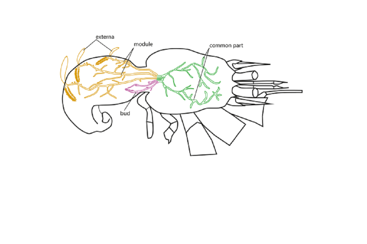
The externa is a sac-like female reproductive organ of parasitic barnacles, positioned outside the host’s body. The externa contains a mantle cavity — a chamber where the developing embryos are brooded.
‘We were interested in exploring how nutrients are transported to the externa, i.e. how nutrients from the interna get to the developing larvae. Previously, several authors described a system of lacunae in the externa — cavities that are connected to the stalks of the interna. The lacunar structures, however, were only described on histological sections. In other words, it was only clear that some cavities exist; yet, the spatial organisation of these lacunae has not been described. We therefore decided to visualise and describe, firstly, the lacunar system of the externa itself, and secondly, the muscular system that might play a role of a propulsatory organ, helping to transport fluid through the lacunar system,’ said Natalia Arbuzova, a master’s student at St Petersburg University.
The zoologists used microcomputed tomography to study the organisation of the lacunar system in parasitic rhizocephala. This method enables visualising various structures in the externa without damaging it. The reconstruction of spatial organisation of different structures in the externa, including its lacunar system, using the tomography method is much easier and visually clearer than using standard histological methods.
Also, the researchers used confocal laser scanning microscopy to study the muscular system. This technique allows for a high-resolution imaging of the muscular system. The structures of interest can be labelled using fluorescent markers, while everything else in the photograph will remain a black background and will not interfere with perception. Additionally, the technique also enables to obtain reconstructions of three-dimensional structures within an object of interest.
The studies using confocal microscopy were carried out in the Resource Centre for Microscopy and Microanalysis of the St Petersburg University Research Park.
‘We suggest that when the circular muscles tighten, the rhizocephalan externa contracts, the lumen of the lacunae narrows and the fluid from the lacunae is ejected into the interna. When the circular muscles relax, the rhizocephalan externa expands, and the lacunae also dilate. Then, after being mixed in the interna, the fluid is pumped into the lacunae of the externa. We have also described the circular muscles at the base of the externa and in the stalks connecting the externa to the interna. Apparently, the circular muscles can close the lumen of the stalk and restrict the flow of liquid either within the externa or from the interna into the externa so that there is time for mixing of the fluid in the interna,’ explained Natalia Arbuzova.
The research was conducted as part the project ‘Morpho-functional role of muscular systems for the evolution of parasitic barnacles (Cirripedia: Rhizocephala)’, supported by a grant from the Russian Science Foundation.
St Petersburg University, the oldest university in Russia, was founded on 28 January (8 February) 1724. This is the day when Peter the Great issued a decree establishing the University and the Russian Academy of Sciences. Today, St Petersburg University is a world-class scientific, educational and cultural centre. In 2024, St Petersburg University will celebrate its 300th anniversary.
The plan of events during the celebration of the anniversary of the University was approved at the meeting of the Organising Committee for the celebration of St Petersburg University’s 300th anniversary. The meeting was chaired by Dmitry Chernyshenko, Deputy Prime Minister of the Russian Federation. Among the events are: the naming of a minor planet in honour of St Petersburg University; the issuance of bank cards with a special design; the creation of postage stamps dedicated to the history of the oldest university in Russia; and the branding of the aircraft of the Rossiya Airlines to name just a few. The University has launched a website dedicated to the upcoming holiday. The website contains information about outstanding University staff, students, and alumni; scientific achievements; and details of preparations for the anniversary.